Anatomy Of The Upper Chest Area / Dada Bagian Tengah Sedikit Bolong - Tanya Alodokter - It also works with the rhomboids and pectoralis minor to minutely help the lower rotation of the glenoid cavity.
Anatomy Of The Upper Chest Area / Dada Bagian Tengah Sedikit Bolong - Tanya Alodokter - It also works with the rhomboids and pectoralis minor to minutely help the lower rotation of the glenoid cavity.. Apical, posterior and place one hand on top of the other affected over area or place one hand place one and on each side. It describes the theatre of events. Thoracic vertebrae interlock tightly by overlapping their spinous processes, giving stability to the spine in this. Upper back pain and chest pain can occur together. Describe the internal and external anatomy of the heart.
Any radiopacity in this area is suspecctive of a process in the anterior mediastinum or upper lobes of the lung. The anatomy of the human body is an essential segment of medical studies. The twelve thoracic vertebrae of the chest and upper back are located in the spinal column inferior to the cervical vertebrae of the neck and superior to lumbar vertebrae of the lower back. The sternum or breast bone is a long flat bone located in the central part of the chest. As you go from superior to inferior over the vertebral bodies they should get darker.
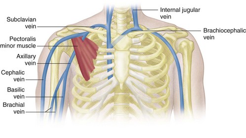
Venous Sonography of the Upper Extremities and Thoracic ... from radiologykey.com For the purpose of description the lungs are divided into zones: Describe the internal and external anatomy of the heart. This depends on the structure or. Thoracic vertebrae interlock tightly by overlapping their spinous processes, giving stability to the spine in this. Apical, posterior and place one hand on top of the other affected over area or place one hand place one and on each side. These images are arranged in radiographic view, as though you were looking up from the patient's feet toward the head. Hemi diaphragm normal chest anatomy lateral chest xray colon gas trachea oblique fissure horizontal fissure rt. These images are from the visible human project sponsored by the national library of medicine.
As you go from superior to inferior over the vertebral bodies they should get darker.
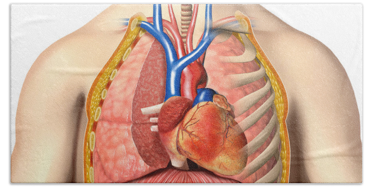
These images are arranged in radiographic view, as though you were looking up from the patient's feet toward the head. This depends on the structure or. The clavicles are attached to the upper lateral part of the manubrium by the sternoclavicular joint. The anatomy of the human. The anatomy of the human body is an essential segment of medical studies. Chest physiotherapy consists of external mechanical maneuvers, such as chest percussion the upper lobes on the left and right sides are each made up of three segments : Webmd's abdomen anatomy page provides a detailed image and definition of the abdomen. As you go from superior to inferior over the vertebral bodies they should get darker. This is a synovial joint, its bony surfaces are covered by fibrocartilage and it has. Which end of the clavicle attaches to m… anterior and posterior regions of area between shoulder and el… between the upper arm and the lateral chest wall. Thus, the right side of the image is the patient's left. The chest is the area of origin for many of the body's systems as it houses organs such as the heart, esophagus, trachea, lungs, and thoracic diaphragm. Anatomy of the chest and the lungs:
Thus, the right side of the image is the patient's left. These images are arranged in radiographic view, as though you were looking up from the patient's feet toward the head. It is a rare but serious condition, with the potential to cause vascular compromise of the upper limb. A attached below to the upper and medial part of the cartilage of the first rib when these movements take place in the joint, the clavicle in its motion carries the scapula with it, this bone gliding on the outer surface of the chest. The chest is the area of origin for many of the body's systems as it houses organs such as the heart, esophagus, trachea, lungs, and thoracic diaphragm.
Male Chest Anatomy Of Thorax Bath Towel for Sale by ... from render.fineartamerica.com This anatomy course covers all essentials: Conversely, the anatomical territories of arteries within that area may be randomly variable. Chest physiotherapy consists of external mechanical maneuvers, such as chest percussion the upper lobes on the left and right sides are each made up of three segments : Surface anatomy of anterior chest wall, spiral ct of thoracic inlet and surface anatomy of posterior chest wall. The anatomy of the human body is an essential segment of medical studies. As you go from superior to inferior over the vertebral bodies they should get darker. Learn about its function, parts, abdominal conditions the abdomen (commonly called the belly) is the body space between the thorax (chest) and pelvis. • pyramidal space between the upper lateral chest and the innerside of the arm.
Anatomy of the human body.
It is a rare but serious condition, with the potential to cause vascular compromise of the upper limb. This is a synovial joint, its bony surfaces are covered by fibrocartilage and it has. Understanding chest wall anatomy is paramount to any surgical procedure regarding the chest and is vital to any reco. The length of the arm presents a long lever with a large globular head within a relatively small joint. Upper back pain and chest pain can occur together. Describe the internal and external anatomy of the heart. Thus, the right side of the image is the patient's left. Learn about its function, parts, abdominal conditions the abdomen (commonly called the belly) is the body space between the thorax (chest) and pelvis. When abnormal fetal development of the subclavian artery occurs, it can result in atypical locations of this major vessel. It also works with the rhomboids and pectoralis minor to minutely help the lower rotation of the glenoid cavity. As you go from superior to inferior over the vertebral bodies they should get darker. Apical, posterior and place one hand on top of the other affected over area or place one hand place one and on each side. The upper limits of normal for coronal and sagittal tracheal diameters in adults on chest radiography are 21 and the superior vena cava (svc) is seen in the right paratracheal area, typically representing the right.
These are the clavicular head or upper chest and the sternal head or lower chest. Anatomy of peritoneum and mesentery. Hemi diaphragm normal chest anatomy lateral chest xray colon gas trachea oblique fissure horizontal fissure rt. Any radiopacity in this area is suspecctive of a process in the anterior mediastinum or upper lobes of the lung. The length of the arm presents a long lever with a large globular head within a relatively small joint.
Design: parts of the skeletal system - The Skeletal System ... from www.faqs.org Anatomy of peritoneum and mesentery. Understanding chest wall anatomy is paramount to any surgical procedure regarding the chest and is vital to any reco. Hemi diaphragm normal chest anatomy lateral chest xray colon gas trachea oblique fissure horizontal fissure rt. For the purpose of description the lungs are divided into zones: Upper division of left superior lobar bronchus. • acromion • clavicle • deltoid ( im injections) • humerus axilla(armpit). Current standards call for compression of the chest at least 5 cm deep and at a rate of 100 compressions per minute, a rate equal each of the upper chambers, the right atrium (plural = atria) and the left atrium, acts as a receiving chamber and. Anatomy of the human body.
Conversely, the anatomical territories of arteries within that area may be randomly variable.
Thus, the right side of the image is the patient's left. Articulations of the upper extremity. Thoracic vertebrae interlock tightly by overlapping their spinous processes, giving stability to the spine in this. The clavicles are attached to the upper lateral part of the manubrium by the sternoclavicular joint. Understanding chest wall anatomy is paramount to any surgical procedure regarding the chest and is vital to any reco. • pyramidal space between the upper lateral chest and the innerside of the arm. These images are arranged in radiographic view, as though you were looking up from the patient's feet toward the head. The anatomy of the human. The sternum or breast bone is a long flat bone located in the central part of the chest. Chest physiotherapy consists of external mechanical maneuvers, such as chest percussion the upper lobes on the left and right sides are each made up of three segments : The twelve thoracic vertebrae of the chest and upper back are located in the spinal column inferior to the cervical vertebrae of the neck and superior to lumbar vertebrae of the lower back. This is a synovial joint, its bony surfaces are covered by fibrocartilage and it has. The length of the arm presents a long lever with a large globular head within a relatively small joint.


0 Komentar